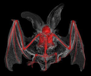
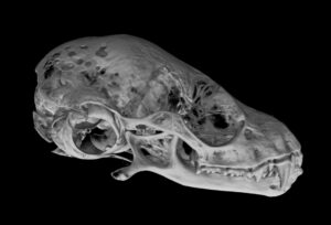
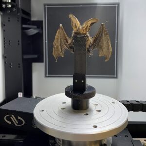
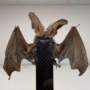
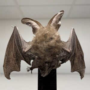
This year to celebrate Halloween, Dr. Andrew Mathers (X-ray CT Project Manager) scanned a brown long-eared bat (Plecotus auritus). X-ray CT enables us to non-destructively visualise the bat’s anatomy in 3D with exceptionally high detail. The various 3D renderings shown, help us visualise not only the bat’s fascinating skeleton but also, its extraordinary anatomical features.
Bats typically use echolocation to find prey, however due to their enormous ears, long-eared bats can sense and hunt prey with the use of hearing alone. This ability is thought to have evolved as a counter measure to various species of moth in turn evolving the ability to hear echolocation frequencies and thus being able to take evasive action when targeted.
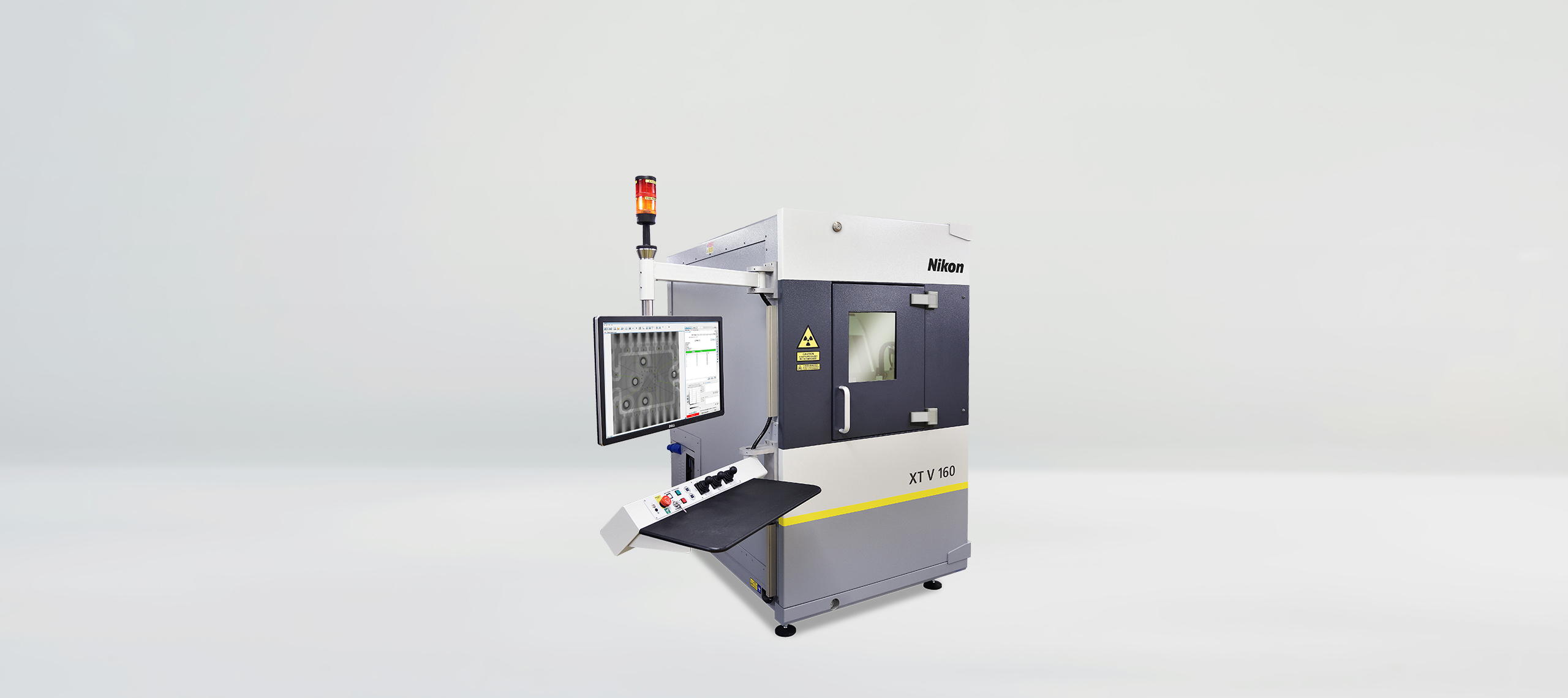
This X-ray CT scan was acquired at a combined X-ray power (Watts) and voxel resolution (µm) of 40, using a Nikon XT H 225 ST 2x. This system comes equipped with up to three different Nikon microfocus X-ray sources: a 180 kV, 20 W transmission target, a 225 kV, 225 W reflection target and a 225 kV, 450 W Rotating.Target 2.0. This X-ray CT scan was performed using the 225 kV reflection target option coupled with a Varex XRD 4343CT flat panel detector. For this scan the detector acquired 4422 projections (individual radiographs) at an exposure time of 5656 ms, resulting in a total scan time of ~7 hours because of the resolution (which is quite a long scan compared to the speed of our X-ray CT scans that can be as low as 1 minute).
X-ray CT data was reconstructed using a modified filtered back projection algorithm in Nikon CT Pro 3D and rendered in Volume Graphics Studio Max 3.5. The 3D images shown are composed of multiple volumes from the same scan rendered using different methods (Isorender, Phong render and X-ray render). These differently rendered volumes are layered on top of one another to create the illusion of movement during flight and the dramatic contrast between the skeleton and body morphology, all in a single image.
