Fossil Day was first established by the U.S. National Park Service in 2010 as part of Earth Science Week, to promote public interest and education in palaeontology. Held each October, Fossil Day encourages people of all ages to discover how fossils reveal the history of life on Earth.
Fossils connect us to ancient ecosystems, long-extinct creatures, and the slow, fascinating processes of evolution and geological transformation. Today, with the help of X-ray CT technology, we can explore these relics more deeply than ever before.
Both palaeontology and archaeology involve the study of delicate, often irreplaceable objects—be they fossilised life forms, ancient tools, ceramics, or skeletal remains. Traditionally, exploring internal structures required physical sectioning or excavation, often resulting in damage or loss.
X-ray Computed Tomography (CT) has changed that. It allows experts to see inside an object, in three dimensions, without even touching its surface.
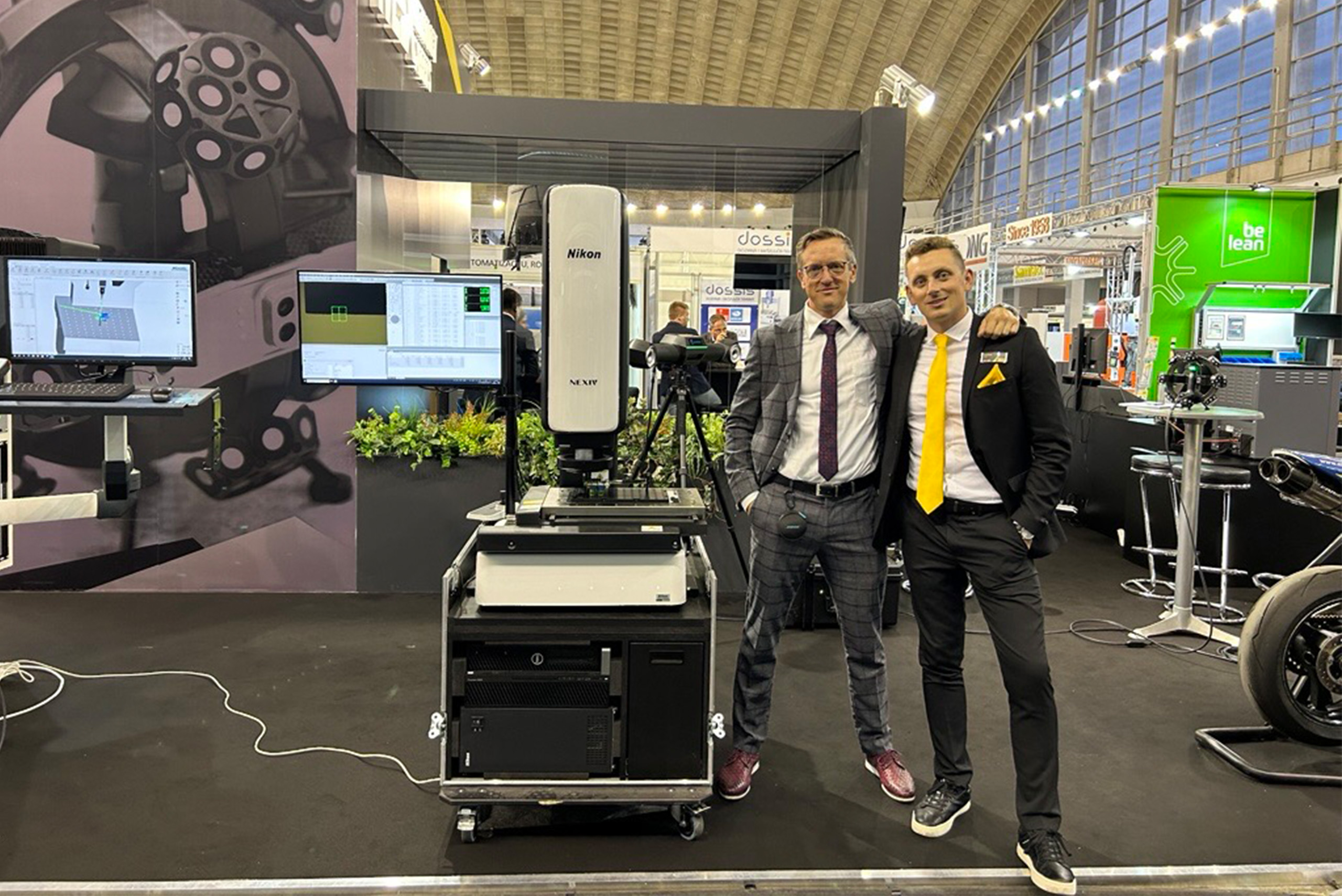
To celebrate this year’s Fossil Day on October 15th, James Finch, Application Engineer X-ray & CT has scanned two fossils that highlight the power of industrial X-ray computed tomography: a beautifully preserved ammonite and a serrated shark tooth.
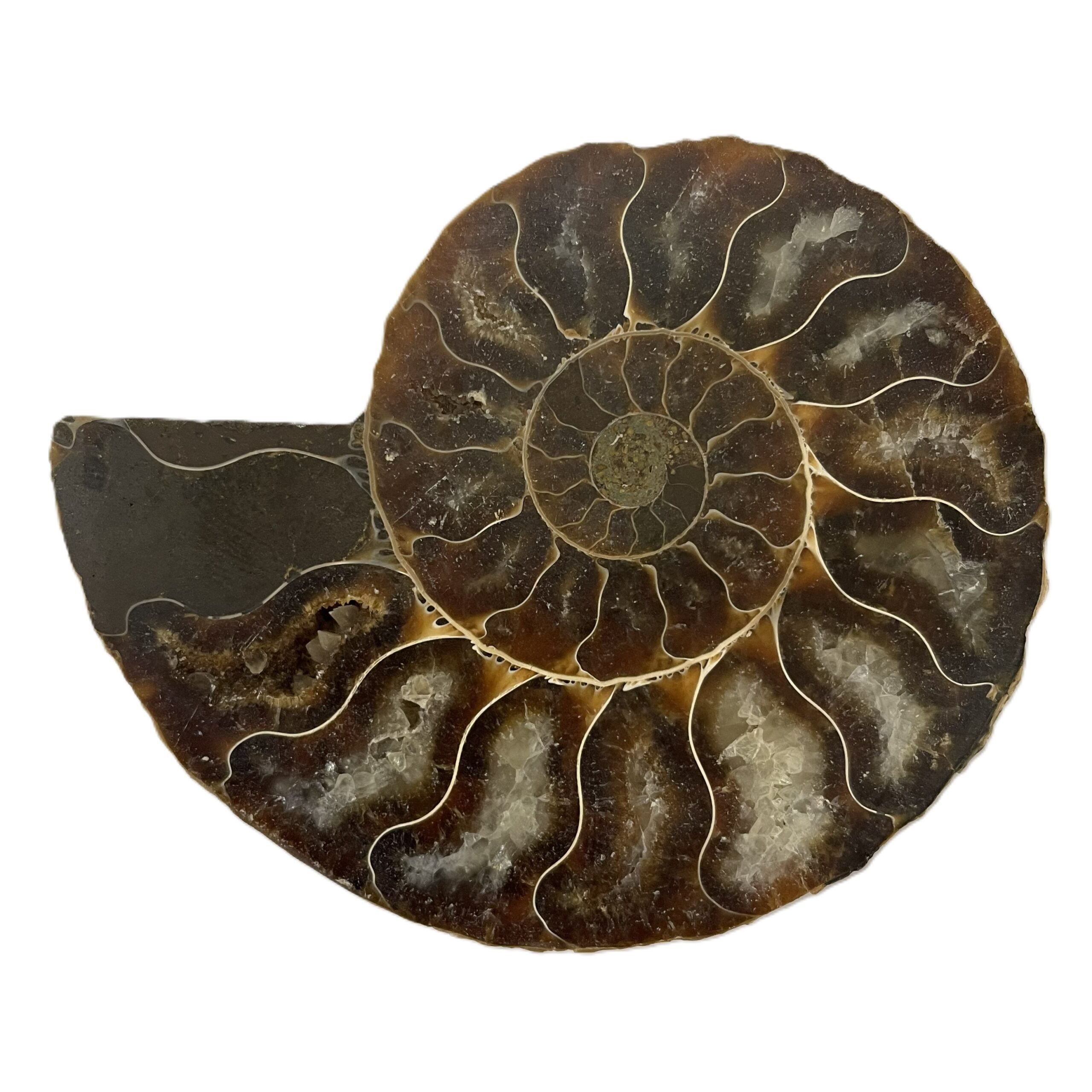
Fossil 1: Ammonite – A Prehistoric Marine Marvel
Ammonites were coiled marine mollusks that lived for hundreds of millions of years before going extinct with the dinosaurs. Their spiral shells, divided into chambers, make them iconic fossils and valuable biostratigraphic markers.
Although the outer shell is often visible, the true complexity of ammonites lies within.
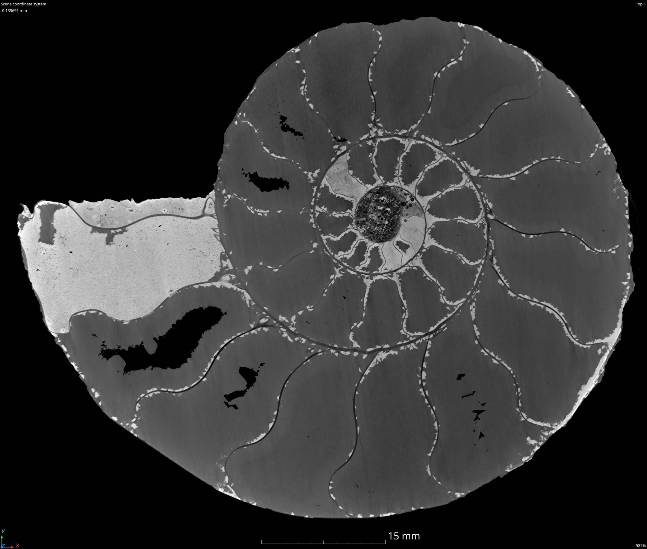
These X-ray CT scans were acquired using Nikon’s Circular CT acquisition algorithm at an X-ray power of 93 W and voxel resolution of 30 µm, using a Nikon XT H 225 ST 2x. The system has one X-ray source, with a choice of multiple targets; 180 kV Transmission Target ; 225 kV static, 225 kV Rotating Target and 225 multi-material target. This scan used the 225 kV rotating reflection target and a Varex XRD 4343CT flat panel detector. The detector acquired 3,546 projections (16 frame/projection) (individual radiographs) at an exposure time of 177 ms and a gain of 6 dB, resulting in a total scan time of 2 hours and 48 minutes.
CT scanning allows paleontologists to study internal chambers, septa, and suture patterns—key traits for classification and functional analysis of ammonites.
Watch the video here:
Fossil 2: Fossilised Shark Tooth – A predator’s legacy
Shark teeth are among the most durable and frequently found vertebrate fossils, with some species shedding thousands of teeth over a lifetime.
The exterior reveals only part of the story. Using X-ray CT, we can detect tooth microfractures, bone density and pathology, porosity changes, and wear patterns—details that can suggest diet, fossilisation environment, or even trauma.
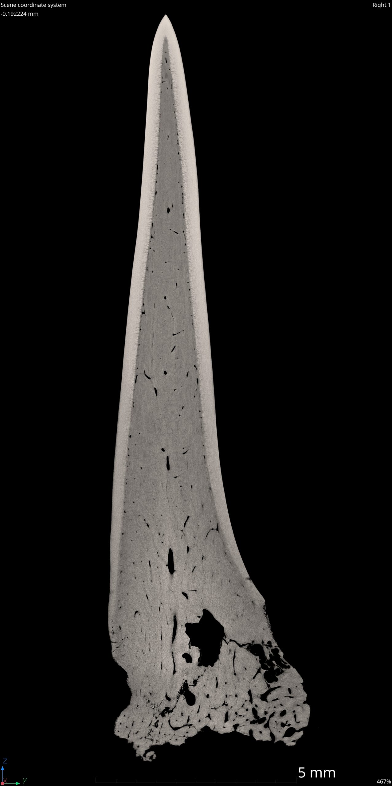
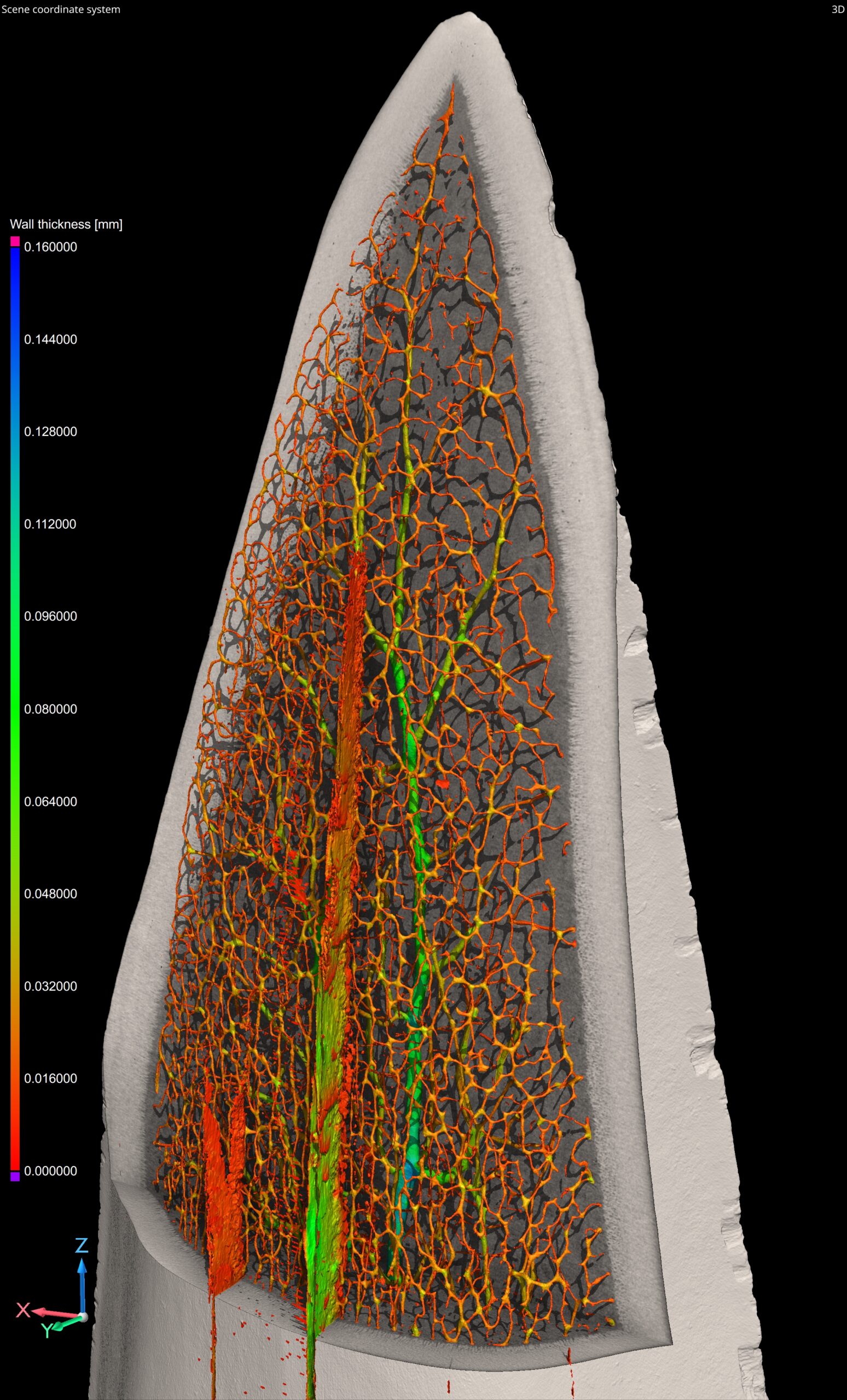
The X-ray CT scans were acquired at 4.15 µm voxel resolution and 7 W, using a Nikon C2 Large Envelope System (LES). This system houses three X-ray sources (a Nikon 450 kV microfocus with rotating target, a Varian 450 kV minifocus, and a Nikon 225 kV microfocus with interchangeable rotating and reflection targets), and two detectors (a flat panel and a Nikon curved linear diode array). For this scan, the Nikon 225 kV microfocus source with Rotating.Target 2.0 and Varex XRD 4343N flat panel detector configuration was used. The detector acquired 7,658 projections (1 frame/projection) (individual radiographs) at an exposure time of 4,000 ms and a gain of 6 dB, resulting in a total scan time of 8 hours and 31 minutes. This is much longer than the normal CT scan of 1-5 minutes, because of the extremely high resolution and complex material.
These high-resolution scans can assist researchers in classifying species, understanding functional morphology, and digitally preserving fossils for further study.
Watch the video here:
Key benefits of Nikon X-ray CT systems for fossil and archaeological analysis
Fossils and archaeological artifacts are often rare, fragile, and embedded in dense materials. Conventional methods like slicing, grinding, or chemical etching can destroy valuable evidence. That’s where X-ray CT offers a significant advantage:
- Non-destructive exploration: Internal features can be visualised without damaging the specimen
- 3D visualization & virtual slicing: Enables precise study from every angle, including features hidden by rock or matrix
- Material density mapping: Identifies mineral infill, voids, and fractures based on attenuation differences
- Digital preservation & sharing: Scans can be archived, replicated via 3D printing, or shared with researchers worldwide
- Quantitative analysis: Enables volume measurements, thickness calculations, and morphological comparisons
- Matrix separation & fossil extraction: CT scans can aid in digital removal of matrix, ideal for fragile or unique specimens
At Nikon Metrology, we’re proud to support researchers, Museums, Universities, and cultural heritage institutions across the globe with advanced CT imaging solutions. From tiny microfossils to large bone fragments, our systems deliver the clarity and precision needed to unlock ancient secrets.
