Nikon’s CT scanning technology has unveiled intricate details of a rare 17th-century pendant watch from the Museum of London’s Cheapside Hoard, transforming experts’ understanding of this unique piece of early modern horology.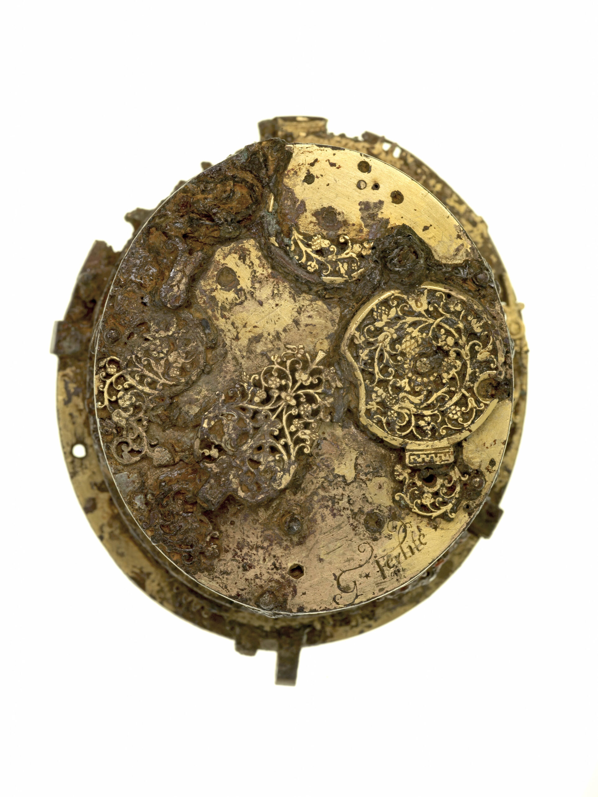
Photo of the movement of the Ferlite watch (Image source: Museum of London).
Industry-leading computed tomography (CT) scanning technology provided by Nikon has been employed to uncover the secrets of a rare 17th-century pendant watch, part of the Museum of London’s renowned Cheapside Hoard.
The intricate details revealed by the scans have transformed the Museum’s understanding of this unique piece of early modern technology, providing new insights into its mechanism, case, and historical significance.
The heart of the goldsmiths’ trade in post-medieval Britain was on the south side of Cheapside in the City of London. Here, on 18th June 1912, beneath a brick-lined cellar floor in a timber-framed building, workmen discovered a remarkable cache of almost 500 items of jewellery, gems and other precious Elizabethan and Jacobean objects.
Probably buried in about 1640 at the onset of the English Civil War, the so-called Cheapside Hoard is now housed in the Museum of London and contains only one item that can be directly attributed. It is a gilt-brass pendant watch, made in Geneva between 1610 and 1620, bearing the signature of its maker, G Ferlite (Gaultier Ferlite).
For a catalogue to be published to coincide with the opening of the new London Museum in 2026-2027, Hazel Forsyth, Senior Curator Medieval & Early Modern Collections responsible for the Cheapside Hoard, wanted to check a few facts and figures concerning recent further research on the watch. In 2023 she approached Nikon for assistance and was delighted when Alistair Watson, X-ray Sales Manager, suggested rescanning the watch.
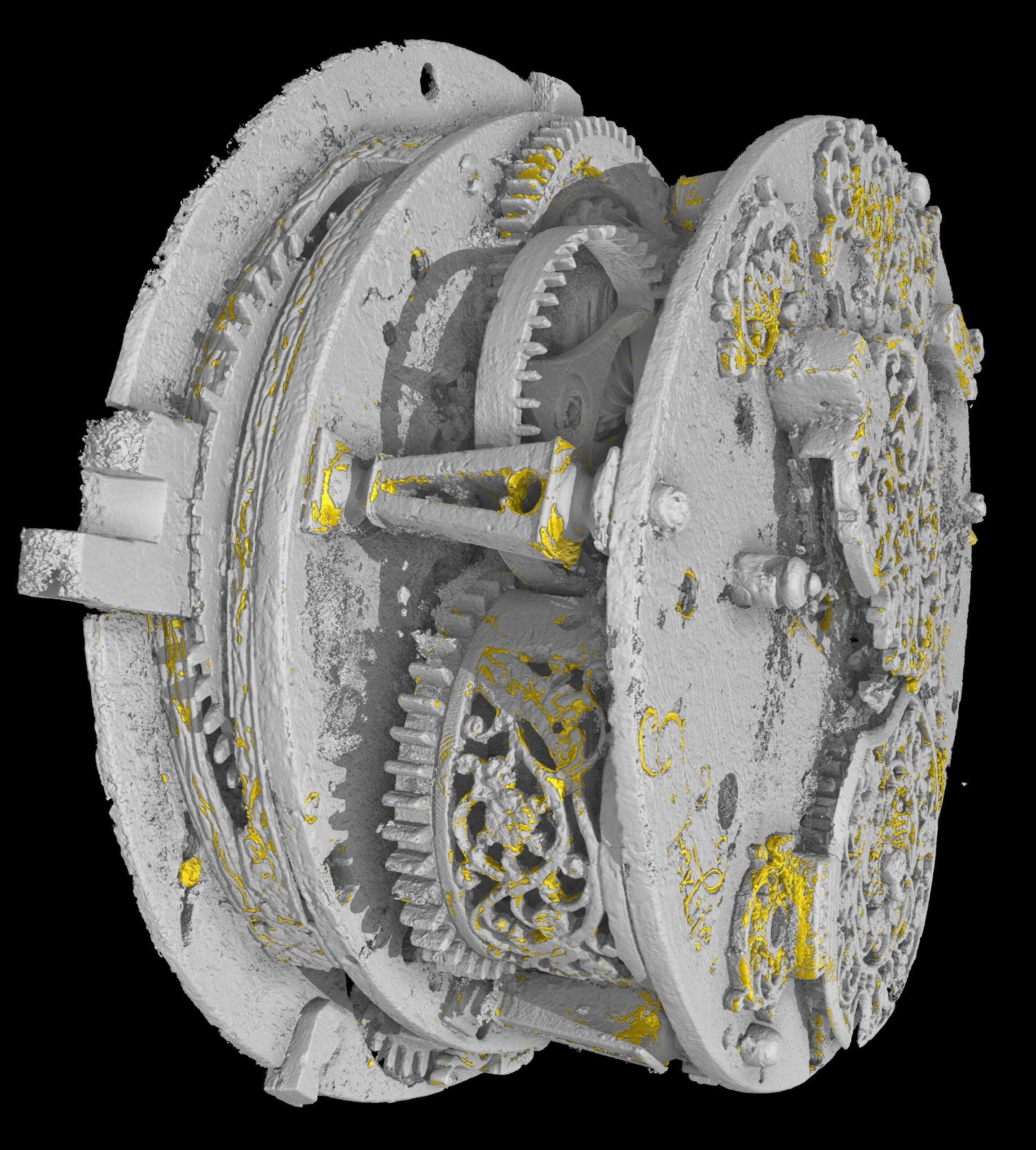
The watch movement had first undergone X-radiography and CT reconstruction in 2005, but the technology has progressed enormously since then and there was potential to discover much more detail. The latest investigation was undertaken by James Finch, Applications Engineer at Nikon’s X-ray CT production, demonstration and subcontract inspection centre in Tring, UK, in February 2024.
The watch was scanned in a Nikon XT H 450 system equipped with a source having a 450 kV rotating reflection target manufactured on-site. Scan parameters were 430 kV, 100 W and a 2 mm tin filter was used. A total of 3,800 projections were acquired with a voxel size of 31.8 microns. Each exposure took 1,415 milliseconds, so the overall scan time was 90 minutes.
Nikon’s CT scans produce “staggering” results
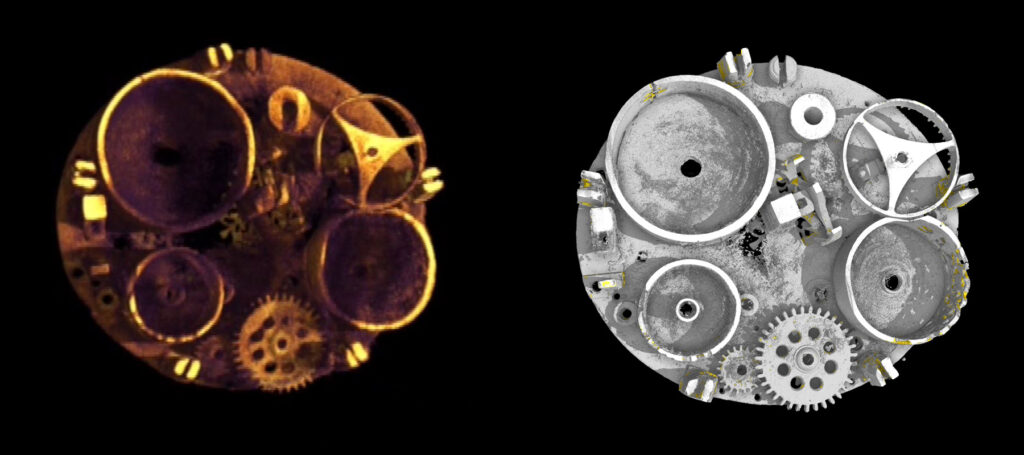
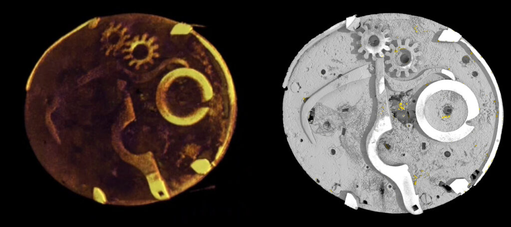
“The results were simply staggering,” says Hazel Forsyth. “For the very first time, it was possible to see clearly details that were blurred in 2005, or simply invisible. The results showed precise features and gear components and relative metal densities, including imaging of the areas which still retain gilding.
“It was also possible to obtain precise measurements of particular features. Even more exciting was the possibility to scan the case as well the movement so that we could virtually re-insert the mechanism into its original housing.
“This meant we could work out what its early 17th century owner would have seen through some of the apertures in dial. The latest images have had a transformative effect on our understanding of this important watch and its place in early modern horology.”
The watch, designed to be worn around the neck, is intriguing because it has a reverse-set chapter ring; the numbers 12 and 6 are transposed so the wearer could read the time by inclining their head. Other interesting features include a piercing in the case for an alarm, as well as astronomical and calendar indications on the dial. When it was made, it would have been a very expensive, luxury item.
Unfortunately, it is in poor condition. Most of the enamel on the dial is lost and the hinge and cover are missing. The dial is currently supported by a modern acrylic insert and the mechanism, which was removed for conservation reasons, is heavily corroded. Very little of the internal structure can be seen with the naked eye, which is why the detail revealed by CT scanning, particularly lately, has been so valuable.
Comparison between the images from the 2024 X-ray CT scans of the Ferlite watch (right) with the scans done in 2005 (left) shows the vast improvement in resolution using the latest technology from Nikon.


