Each year on March 3rd, the world celebrates World Wildlife Day, highlighting the importance of protecting biodiversity and deepening our understanding of the species that sustain ecosystems. For this March edition of Scan of the Month, we focused on one of nature’s most vital pollinators: the bumblebee. Small in size but immense in ecological importance, bumblebees play a critical role in supporting both wild plants and agricultural crops. To better understand their remarkable biological design, Benoit Guilleux, X-ray Project Engineer, used high-resolution X-ray CT to reveal the intricate internal architecture of this delicate insect in extraordinary detail, entirely non-destructively.
Hidden marvels of the bumblebee
Beyond their role as pollinators, bumblebees are remarkable for their indirect flight muscles, which allow them to beat their wings at very high frequencies without directly contracting the wing muscles. This enables them to generate lift beyond what their body size would suggest, a fact that once led scientists to joke that “bumblebees shouldn’t be able to fly.” The combination of thoracic muscles, flexible wing joints, and air sacs forms an efficient biomechanical system, perfectly adapted for hovering, agile maneuvers, and long flights.
Exploring nature’s micro-engineering
Imaging biological specimens presents distinct challenges. Organic tissues typically offer low X-ray absorption contrast, while their structural features can be exceptionally fine. Achieving meaningful visualisation therefore requires carefully optimised parameters that balance resolution, contrast, and scan stability.
Nikon’s X-ray CT technology addresses these challenges through precise control of voltage, power, detector sensitivity, and geometric magnification. For low-density, delicate samples such as a bumblebee, lower accelerating voltages and carefully adjusted power settings enhance material contrast while preserving fine structural fidelity. Detector gain and exposure time can be optimised to maximise signal-to-noise performance, enabling subtle anatomical features to be resolved at micron scale.
Importantly, this flexibility highlights the versatility of Nikon systems. The same XT H 225 platform used to capture this fragile biological specimen can also be configured for high-density industrial applications, such as scanning rock cores or metal castings. With a rapid change to a rotating target and adjusted energy settings, the system can penetrate dense materials while maintaining measurement accuracy and inspection efficiency.
This adaptability ensures that a single X-ray CT system can move seamlessly between delicate natural history specimens and demanding industrial components, delivering high-quality results across an exceptionally broad range of applications.
Capturing the details
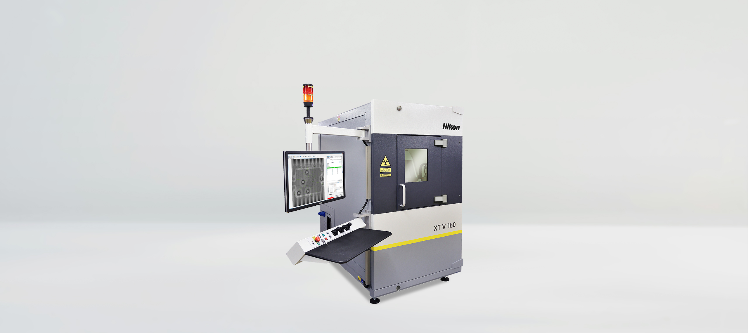
The bumblebee was scanned using the XT H 225 ST 2x system, equipped with a 225 kV multi-metal Static Reflection target with a silver target. For this application, the system was operated at 60 kV and 7 W, a configuration chosen to enhance contrast across low-density biological structures while maintaining fine structural fidelity. Data was acquired using a Circular CT scan technique in combination with a Varex XRD 4343CT detector.
To capture the internal features of the specimen, the scan was performed at a voxel resolution of 7.4 µm. A gain setting of 12 dB and an exposure time of 8000 ms per projection ensured sufficient signal-to-noise performance for the delicate anatomical details. In total, 1200 projections were acquired with one frame per projection, resulting in a complete scan time of 2 hours, 40 minutes and 48 seconds. Please note that this detailed work required a scan longer than the typical X-ray CT scans performed by Nikon technology, which can be as low as 1 minute with a configurable number of projections.
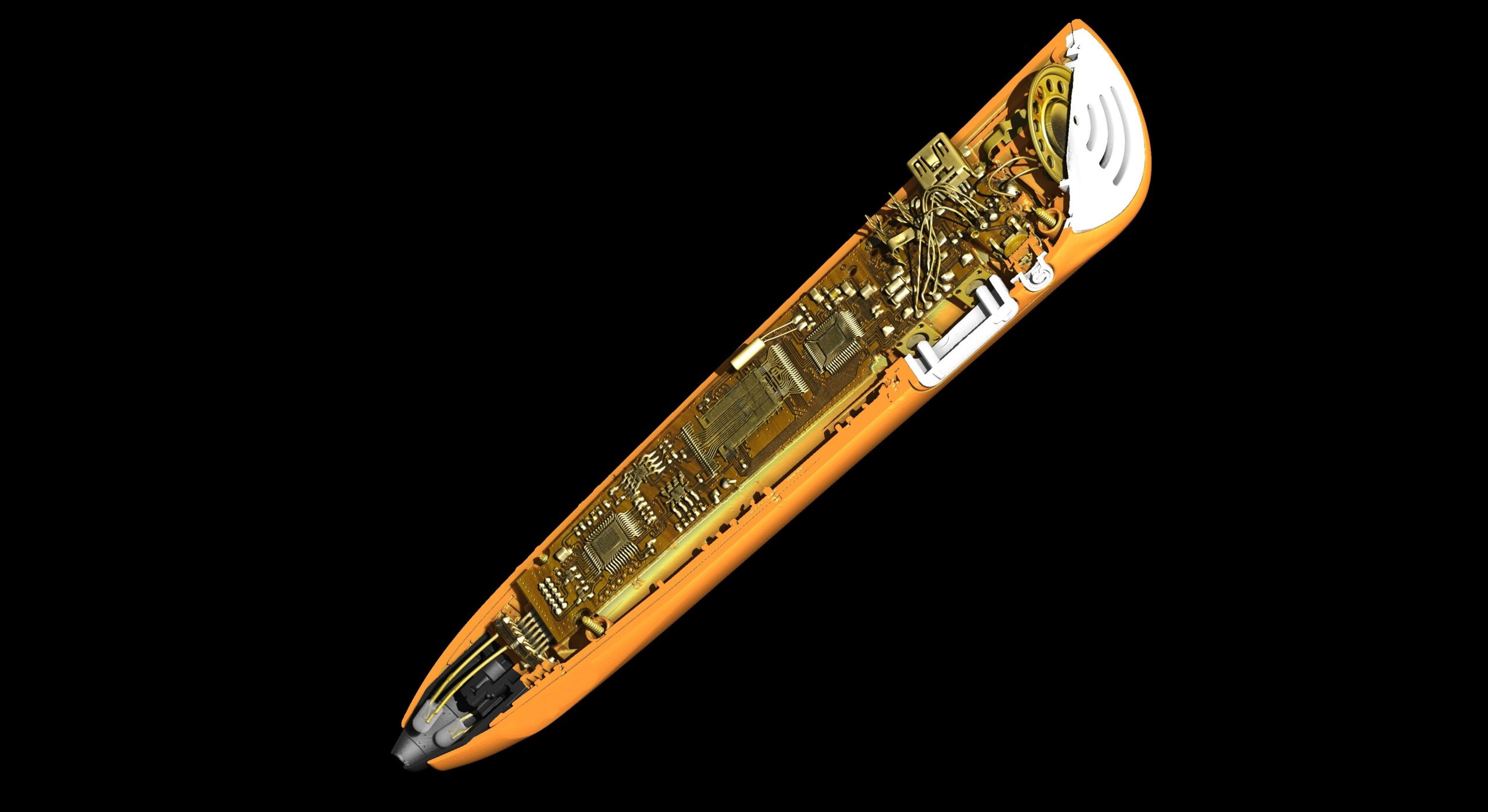
From thorax to wing joint, detail at micron scale
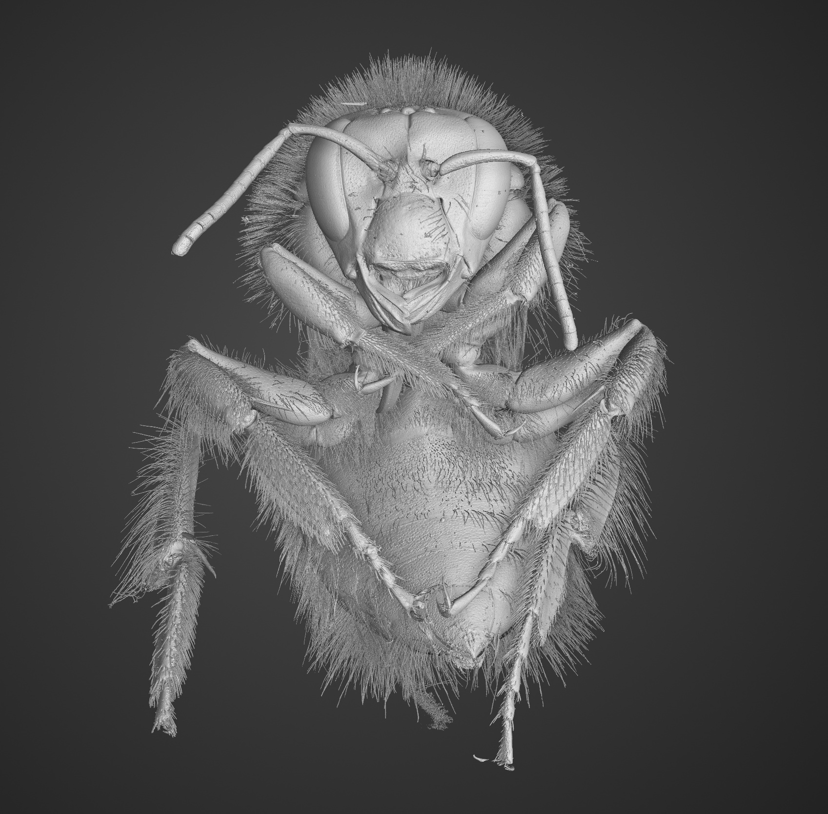
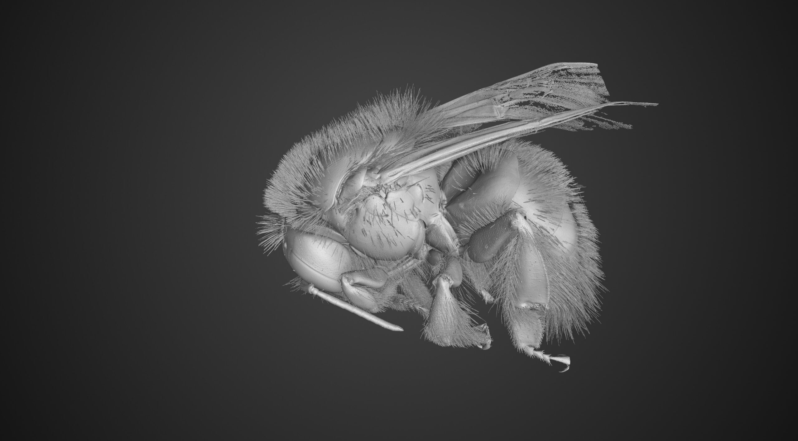
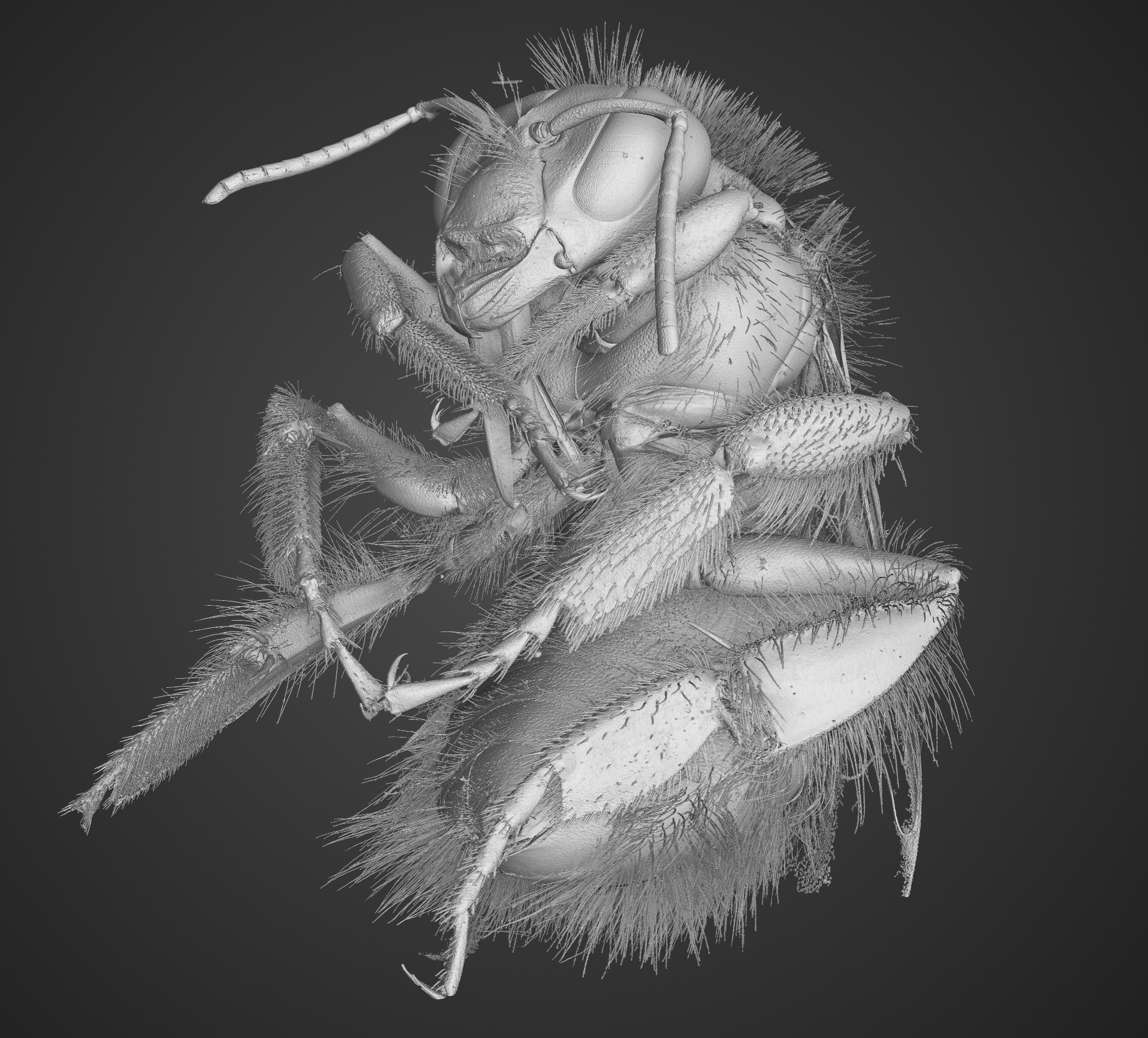
The reconstructed volumetric dataset reveals the extraordinary internal complexity of the bumblebee. Within the thorax, dense indirect flight muscles, responsible for the insect’s rapid wingbeats, are clearly distinguishable. The segmentation also highlights variations in exoskeletal thickness, fine joint geometries at the wing articulation points, and internal air sac structures that contribute to flight efficiency.
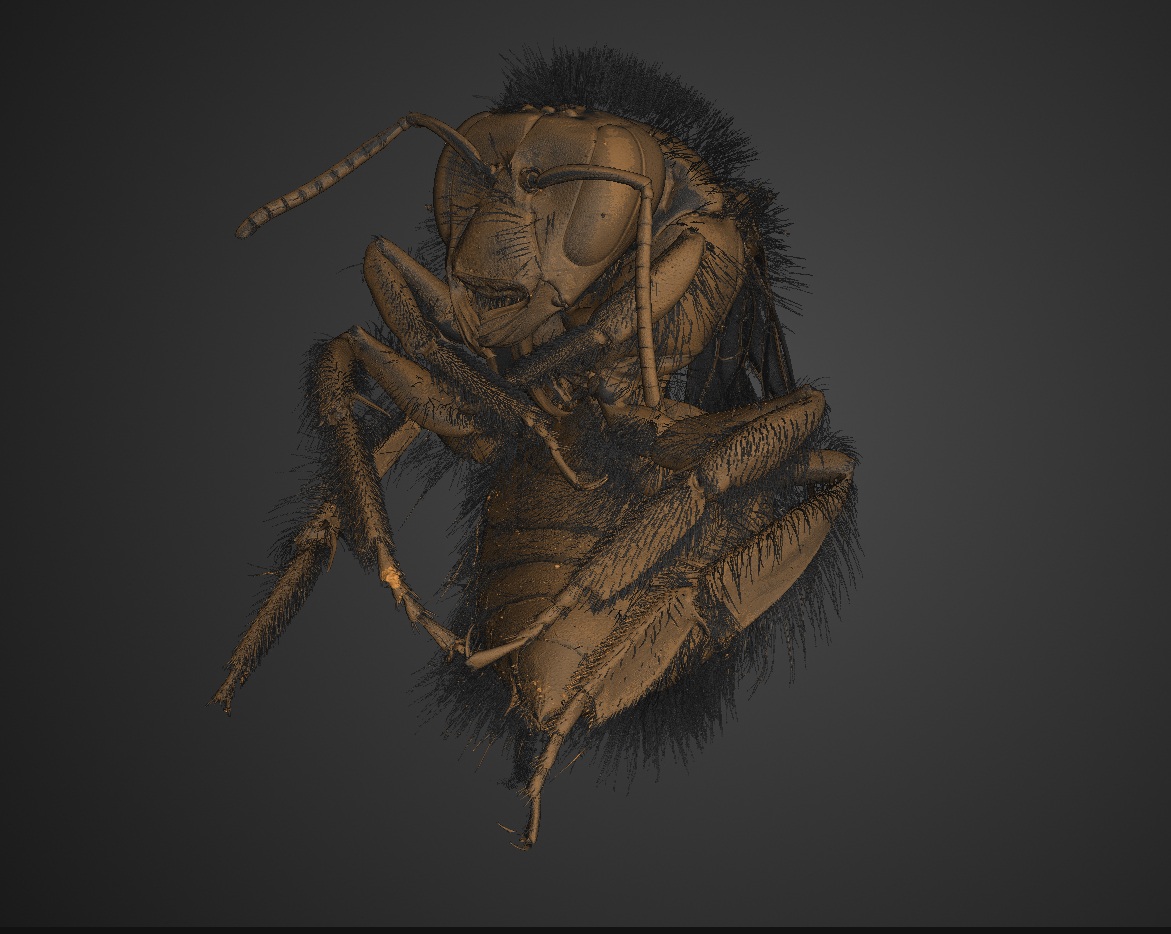
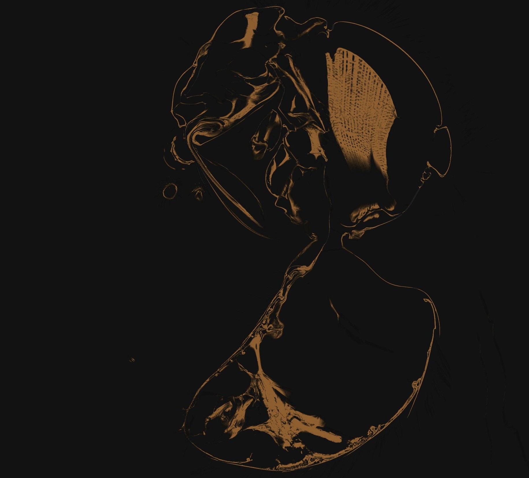
At a voxel size of just 7.4 microns, even subtle structural transitions become visible, enabling both qualitative visualisation and quantitative analysis. Researchers can examine morphometrics, assess structural symmetry, and digitally preserve the specimen without any physical sectioning.

Precision technology in service of Biodiversity
On World Wildlife Day, this scan of a single bumblebee demonstrates that even the smallest organisms have remarkable structural complexity. Nikon Metrology’s advanced imaging solutions allows researchers to examine these details non-destructively, supporting scientific analysis, digital preservation, and a deeper understanding of the natural world.
