CASP, a charitable organisation supporting energy transition through geological research, relies on Nikon’s SMZ18 research stereomicroscope for in-depth rock samples and fossils analysis. The advanced microscope’s versatility and precision have made it an essential tool for CASP scientists, enabling them to contribute to sustainable energy solutions.
Nikon, a pioneer in advanced microscopy, has played a pivotal role in supporting the Cambridge Arctic Shelf Programme’s (CASP) vital geological research for energy transition, thanks to almost a decade of work with Nikon’s SMZ18 stereomicroscope.
The Nikon SMZ18 has become a cornerstone of CASP’s research infrastructure, serving the needs of scientists, visiting researchers, and students. Its versatility allows for diverse applications, from studying the surface details of fossils to extracting zircon grains for radiometric dating.
CASP is a not-for-profit, charitable organisation conducting geological research on rock samples from outcrops and well cores to support energy transition. Building on several decades of research experience in sedimentary basins, CASP primarily aids the characterisation and assessment of geological CO2 storage targets, aligning with national and international Net Zero strategies.
The organisation was formed in 1948 when W. B. Harland launched the Cambridge Spitsbergen Expeditions (CSE). Many senior figures in academia and industry gained early field experience on these expeditions. CSE gave rise to the Cambridge Arctic Shelf Programme in 1975 and CASP was incorporated in 1988 with charitable status.
Subsequently, CASP greatly increased its regional scope, diversifying its research to areas outside the Arctic, and forging a reputation for conducting safe, high-quality, field-based geological research in frontier and underexplored basins. This work was supported by multinational hydrocarbon companies, primarily to improve their geological knowledge of new areas.
Microscopy key to analysis of CASP’s core collections
Today, the core of CASP’s work is collecting new geological data. This involves detailed descriptions and photographic documentation of rocks in the field or core repositories, as well as the collection of samples for analytical work back in Cambridge.
In the CASP laboratory, microscopy is a key element of the research process to characterise and categorise the collected samples. Most of CASP’s current projects investigate the CO2 storage potential of rocks in the Southern North Sea. Simon Schneider, who holds a PhD in palaeontology from Ludwig-Maximilians-University, Munich, is leading one of these projects.
Dr Schneider explains: “Despite the recent acquisition of an advanced Scanning Electron Microscope, optical microscopy remains an essential analytical technique. The samples we analyse are often only a few centimetres in size, and we are usually interested in their composition and, thus, in much smaller objects or structures within. Most commonly, we look at rock thin sections, which are 30 to 70 microns thick, semi-translucent slices of rock glued on a thin 30 x 60 mm size glass slide.
“To view rock fragments, mineral grains or fossils satisfactorily under a microscope, we need a range of different magnifications and, importantly also, a reasonable working distance. Previously, we had a couple of older binocular microscopes on which the transmitted and incident lighting was less than satisfactory. These instruments also often took a long time to set up.”
The alternative was to utilise a powerful Nikon monocular microscope that has been in use at the laboratory for many years, but its x200 to x2,000 magnification range is too high to allow CASP scientists to assess larger samples and to characterise easily the mineral or fossil content of the rocks.
The latter instrument was fitted with a Nikon digital camera for the microscope, so when CASP looked for a more efficient, lower-power stereomicroscope for their needs, the prospect of a model from the same manufacturer was attractive, as the same camera could be used on both microscopes. Nevertheless, several different makes were reviewed before a Nikon SMZ18 research stereomicroscope was purchased from Nikon in Derby, UK.
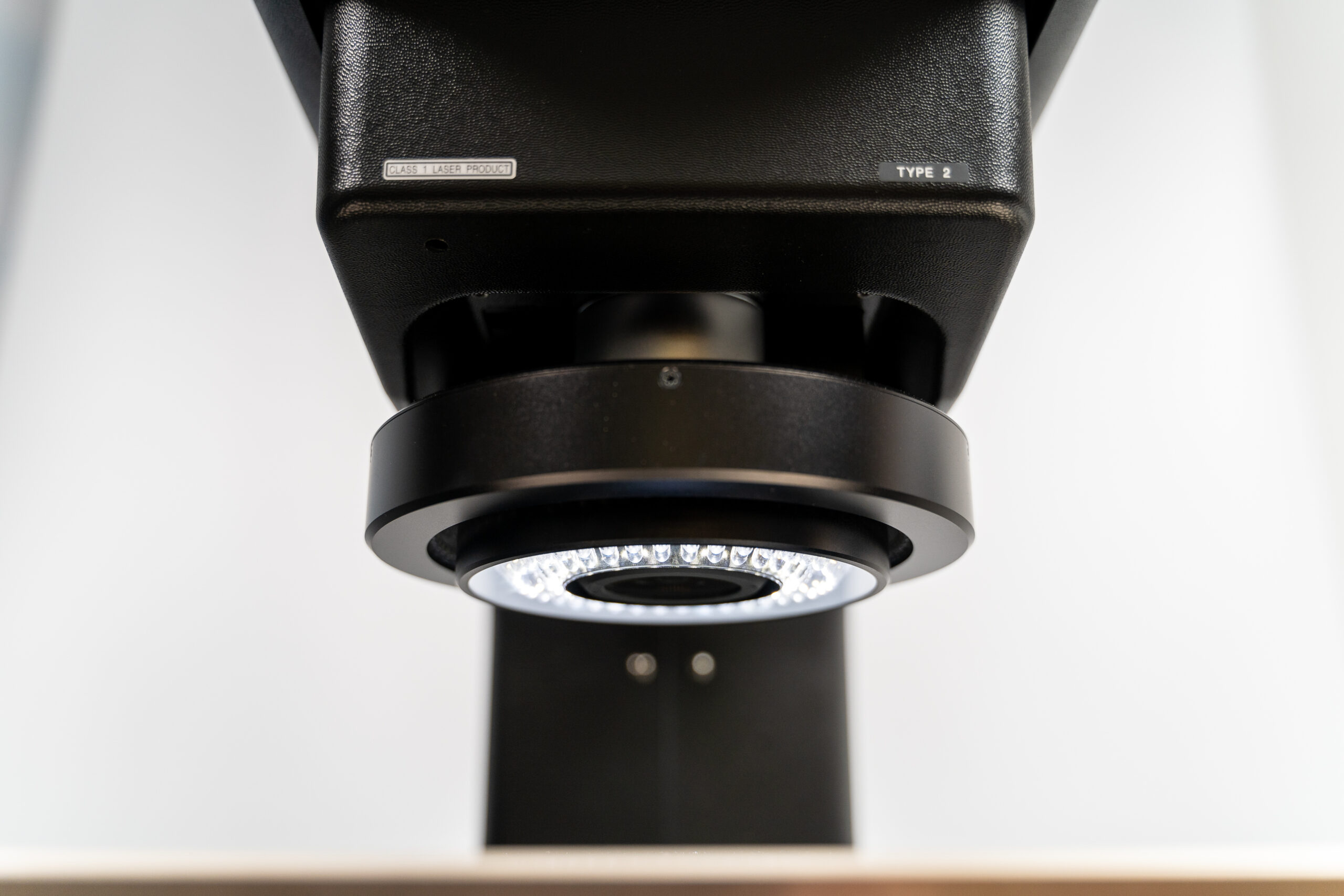
The microscope has macro and micro imaging in one manually controlled instrument to conveniently view and manipulate samples. A high-performance lens provides clear images with uniform brightness across the entire field of view.
Nikon’s world-renowned optics perfect for CASP
Dr Schneider confirms: “The Nikon SMZ18 is a brilliant microscope. It has 18:1 manual zoom, and our binocular eyepiece provides a 22 mm field of view. The magnification range up to x270 and the 60 mm working distance of our objective are perfect for what we need. The quality of the optics is world-renowned. Another crucial factor is the effective episcopic [reflected light] and diascopic [transmitted light] illumination of our samples.”
Apart from studying rocks, Simon Schneider often uses the microscope to look at the surface details of fossils, usually shells. However, he is not the only researcher at CASP who has benefited from the Nikon SMZ18 research stereomicroscope. Over nearly nine years, the Nikon SMZ18 has been regularly used by CASP employees, visiting scientists, and students.
Drs Michael Flowerdew and Michael Pointon specialise in geochronology and geochemistry and routinely use the instrument to extract zircon grains from mineral samples for radiometric dating purposes. Another regular user is Dr Simon Passey, who primarily studies volcanic rocks and their geochemical interaction with sandstones.
The composition and texture of sandstones is also the main research topic of Dr Michelle Shiers. The Nikon SMZ18 has served the needs of scientists well for nearly a decade and continues to be a powerful piece of CASP’s research infrastructure.
Explore the full range of Nikon’s Industrial Microscopy here.

