Industrial X-ray equipment from Nikon Metrology “just works and works with very little fuss, even after seven years”, allowing Norwegian flora to be seen in a new light – literally.
It is 250 years since Norwegian bishop and botanist Johan Ernst Gunnerus published a book in Latin, “Flora Norvegica”, about plants in his native country. In 2020, professor Øyvind Hammer from the Natural History Museum at the University of Oslo and his scientific researcher and botanist wife, Marte Holten-Jørgensen, paid homage to the highly skilled natural historian by entitling their new book on Norwegian flora, “Flora Norvegica Radiographica”. It took three years to write and is published by Spartacus Forlag.
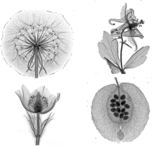
As its name implies, the book is a compilation of X-ray images of plants, 110 in all showing in stunning detail the interior of many species of flora found in urban environments, by the roadside and in the wild. Each image is accompanied by a descriptive text detailing how the plant lives as well as its place in medicine, folk lore and literature. Rather than a scientific textbook, the publication is an unrestrained celebration of the diversity, beauty and significance of plants for the human race. Most are everyday species such as the dandelion, maple, lily of the valley, white willow, wild strawberry, gossip cabbage and wrinkled rose, while others are rare and endangered. Prof Hammer said, “My job frequently involves me X-raying all kinds of items including fossils, artefacts and insects, as well as flowers and plants although I was never really so interested in them until the radiographs opened my eyes to how beautiful and exciting plants in Norway really are.
“Then I looked on the internet and saw there is a long history of taking X-ray images of plants; it has been seen as a sort of underground art form for more than a century. The new book is a side-project, but it fits with my job as a professor in the museum by shining a light on and promoting Norwegian nature.”
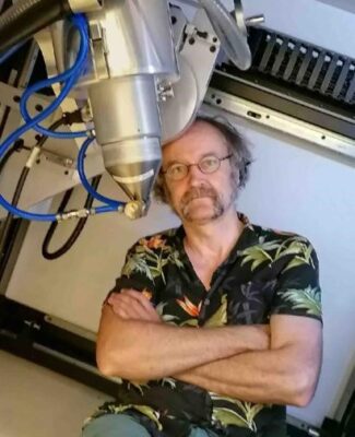
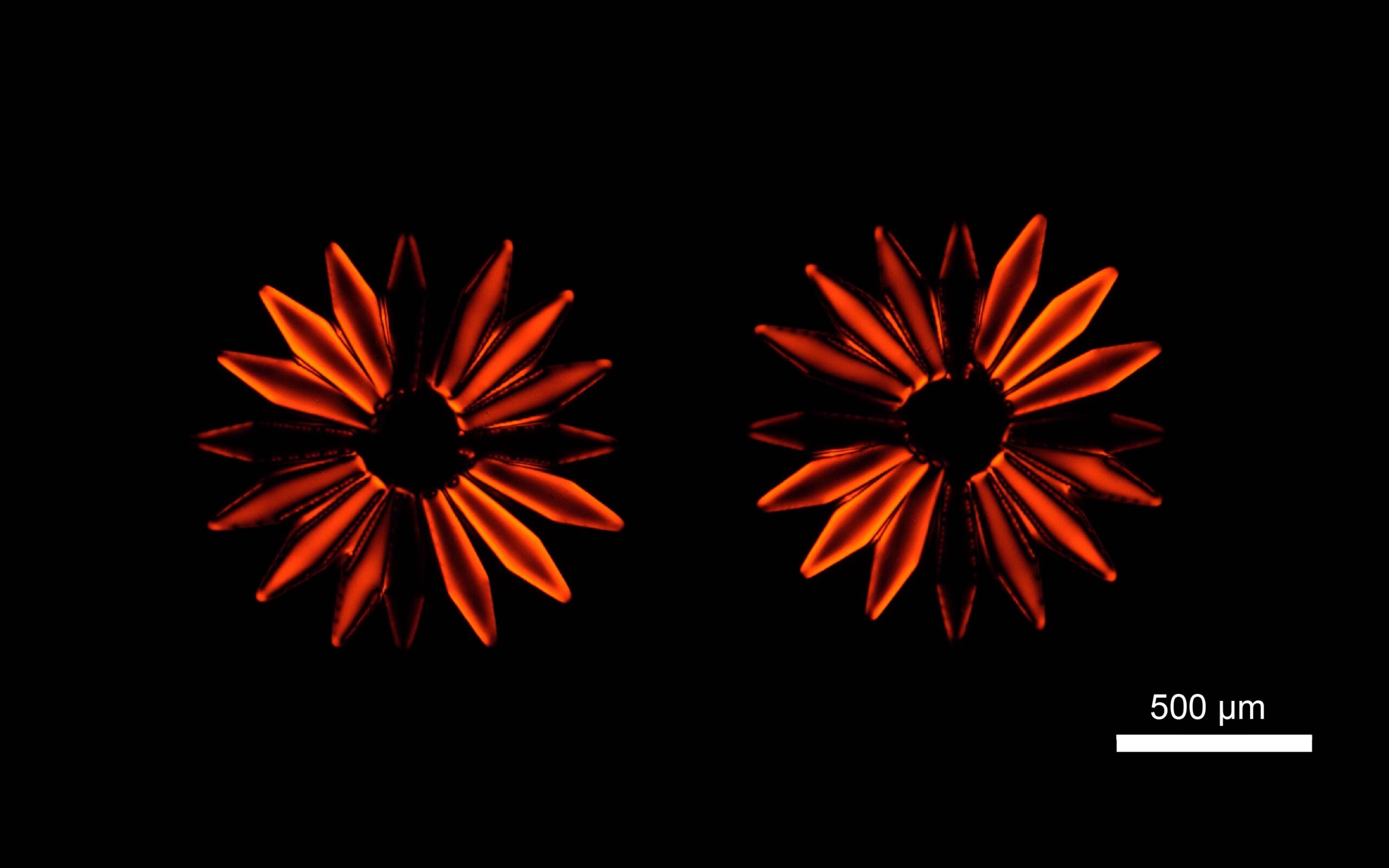
The light, of course, is from the X-ray part of the electromagnetic spectrum, rather than the visible frequency band, and in this case is generated in an XT H 225 ST X-ray CT (computed tomography) system supplied to the museum seven years ago by Nikon Metrology. It is Prof Hammer’s favorite piece of equipment and he was responsible for producing all the images, which were made using a microfocus transmission target so even the smallest details may be seen. Ms Jorgensen wrote the book and they both shared the foraging and collection of samples.
Up to 300 images were made, each involving around three hours’ work including finding and picking the plant, mounting it in the X-ray scanner, producing the image by taking multiple X-rays and averaging them, and then post processing the data. There was no time pressure during this project to achieve results quickly, so Prof Hammer took advantage of the flexibility of the Nikon Metrology system to choose instrument settings that extended the time taken for data acquisition and integration of the images so that optimal images could be acquired.
He added, “The X-ray machine makes us see the diversity and fragile beauty of plants in a new light, literally. Both scientifically and aesthetically, the images are absolutely fabulous and many of them are razor sharp. I think my main emotion is an enormous enthusiasm for the beauty of X-ray and CT images in general.”
“The smooth and consistent performance of the Nikon Metrology instrument in particular made the job easy. It just works and works with very little fuss, even after seven years. Our next literary project will be to write a book on the rich sealife around the coast of Norway.”
Geologists, biologists and archaeologists are the other major users of the X-ray machine at the Natural History Museum in Oslo. X-ray CT is especially useful for studying fossils, according to Prof Hammer, as it allows researchers to see inside a rock and observe the fossil within it in 3D, without destroying the specimen.
Interestingly, when X-raying plants for the book, he chose not to produce 3D computed tomograms from the X-rays but to keep the images in two dimensions. The density and thickness of the various elements in the plant determine how many of the X-rays penetrate through to the detector and therefore the shade of grey in the monochromatic image. It enables very fine detail to be seen inside the flower, such as nectaries and seeds, whereas normally they would be obscured by leaves and other external structures. It is analogous to the old method of producing a herbarium, but instead of being flattening and therefore distorted the specimens remain lying or hanging in their natural shape, so the record is more accurate.
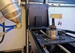
XT H 225 ST installed in the Natural History Museum at the University of Oslo
In 2013 the University of Oslo purchased a Nikon Metrology XT H 225 ST with both a transmission target and a reflection target for maximum flexibility when scanning small, soft objects and large, highdensity objects. The Natural History Museum has been using the instrument extensively for investigating a wide variety of objects, including sediment cores, rock cores (up to 10 cm in diameter), fossils, insects, skeletal material, mineral samples and samples for physics experiments. Two sources are used in the system: – 180 kV transmission source with 1µm spot size – 225 kV reflection target has a minimum spot size of 3µm and a multi-metal target allowing rapid change of target material (W,Cu,Mo,Ag) Prof Hammer uses the Nikon XT H 225 ST system for researching rocks and fossils. His main passions are cephalopods and other invertebrates as well as foraminifera. (And now also plants and flowers!)

