
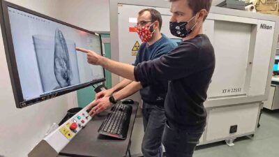
The Natural History Museum (NHM) in London is one of the few museums in the world to automate the X-ray CT scanning of specimens. It recently took delivery of its second CT scanner from Nikon Metrology, part of the global Nikon Corporation, which has extensive experience working with and advising museums on every continent.
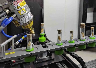
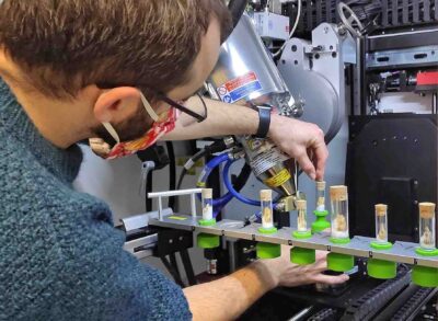
The latest Nikon XTH225 ST system is fitted with a 10-station sample autoloader whereby racks of samples are loaded inside the cabinet, enabling the system’s in-built manipulator to automatically load them onto the scanning turntable and subsequently retrieve them.
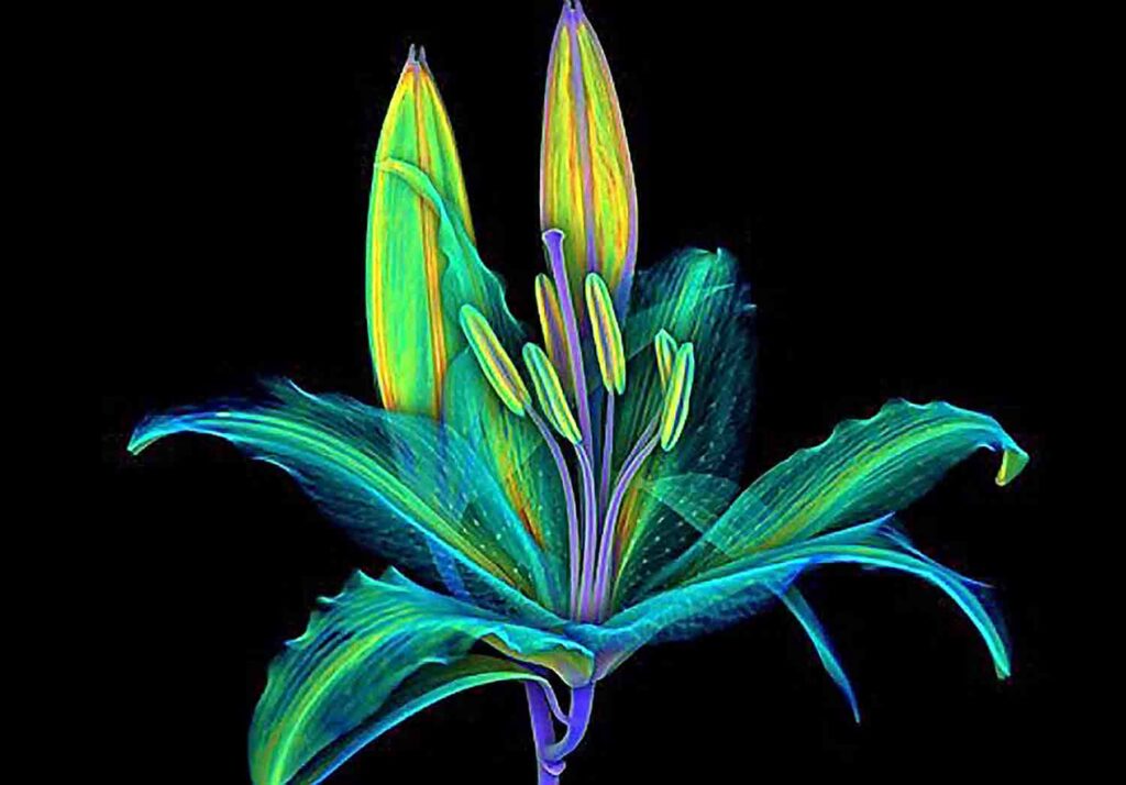
The purpose of the initiative at the NHM is not only to assist the Museum’s researchers across its many departments and disciplines, but also to speed up the non-destructive, non-invasive inspection and digitisation of its vast collections, including artefacts, animals, plants, fossils, rocks and meteorites.
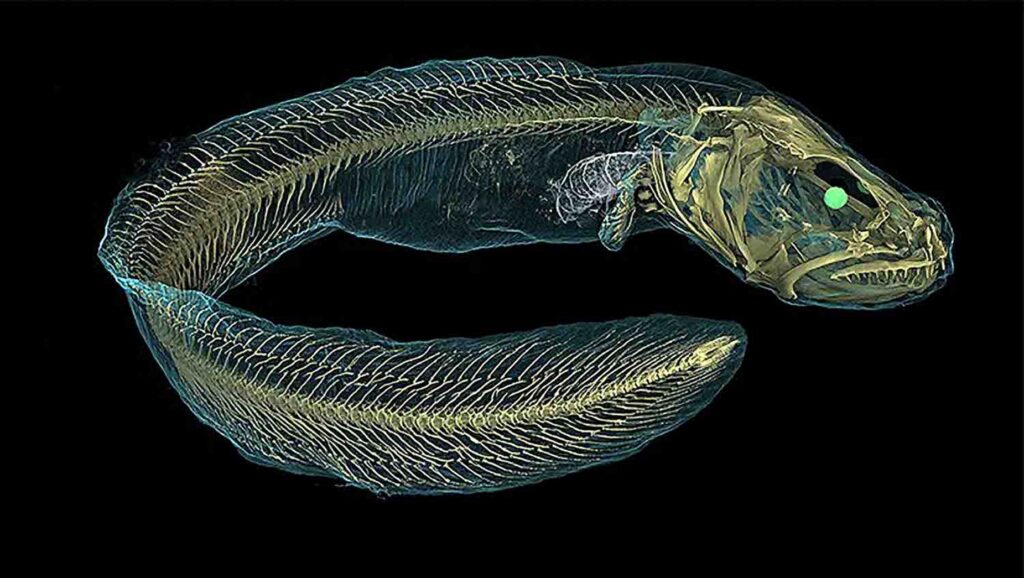
Many types of animals undergo investigation, including for morphometric comparison of their skeletons. It is an easy way to obtain accurate, repeatable results for computer analysis to assist in the study of evolutionary trends, without introducing the variability of human observation. For example, corals (which although immobile, are classed as animals) are routinely scanned as part of studies into global warming.
Additionally, the XTH225 ST is used by industrial customers for non-destructive 3D analysis of samples such as batteries, parts produced by additive manufacturing, and honeycomb structures. Industrial applications account for about 20% of the system’s throughput and contribute the Museum’s annual revenue.
The data acquisition process is accelerating the ongoing imaging of the NHM’s collection of specimens so that the database can be populated faster with 3D digitised models of both
 their external and internal features. The result is that information may be shared more quickly with researchers, conservators, academia, business and the general public internationally.
Being able to use the instrument’s own handling equipment to automatically load items for scanning was a key driver in our decision making for purchasing this micro-CT scanner. -Dr Vincent Fernandez, micro-CT laboratory manager at the Museum
“We also recognise the need for reliability of the equipment and its imaging quality. The Museum’s system is often running 24/7. Our previous scanner, a Nikon 225 kV micro-CT system delivered back in 2008, is still in operation today at an industrial company on the continent.”
He further advised that the automation equipment at the NHM is used extensively for unattended inspection of items small enough to sit on each of the ten sample support stations in the rack, each of which is approximately 5cm in diameter.
With any CT system, long scan times provide the best results. This is where the sample autoloader excels, allowing unattended scan times of up to two hours per sample overnight and at weekends, maximising the throughput while providing the best possible resolution. However, if maximum resolution is sacrificed for higher speed and throughput, a typical requirement for industrial applications, the system is capable of 3D scanning and reconstruction in under two minutes.
The XTH225 ST has a maximum field of view of 42.75 x 42.75 cm, or 85.5 x 85.5 cm using offset CT, and can accommodate samples up to 70 cm high. These larger samples are mounted directly on the turntable manually and the taller samples are digitised by stitching multiple scans, or using the X.tend helical scan software to produce a single extended image with no vertical cone beam artefacts.
Scanning productivity is raised not only by the autoloader for smaller samples, but also by Nikon’s latest, unique, Rotating Target 2.0. The power that can be put into a static target is limited by the material’s heat dissipation; too much power will melt the target, resulting in loss of focus. Nikon’s rotating target overcomes this by spinning a water-cooled target at 6,000 rpm, thereby dissipating the heat over a peripheral track and allowing either the X-ray power or resolution to be trebled. The rotating target can be used for greatly increasing scanning speeds or for producing a smaller focal spot size for a given power compared to a static target.
The rotating target can run continuously at 450 watts without requiring any cool-down period. This power combined with the latest 8 megapixel, 16 bit detector allows data acquisition rates of 15 fps full resolution, or 30 fps with the detector binned 2 x 2 pixels.
For the above reasons, Dr Fernandez keeps the rotating target in place for most of the time the system is in use. However, if a smaller focal spot size is required, the system has been supplied with an easily interchangeable, 3-micron static target and a 1-micron transmission target for even greater resolution. Dr Fernandez describes the transmission target as “excellent for studying very small samples”.
Additionally, the system is supplied with Nikon’s unique multi-metal target. Besides the standard tungsten target, the operator can select three alternative materials, molybdenum being the second most useful as it is ideal for studying delicate specimens like a leaf or a plastic component. There is also a choice of silver and copper targets for different applications and all may be exchanged without breaking the vacuum of the source tube.
The equipment is easy to use and allows us to finely adjust settings for optimally studying a wide range of different objects. With the added advantage of automated loading and unloading of specimens, we have been able to scan an average of 100 items per month since we started operating the new micro-CT system.


