This month, Dr. Andrew Mathers (X-ray CT Project Manager) scanned a 70mm long, common UK lizard (Zootoca vivipara).
X-ray CT enables us to non-destructively visualise the lizard’s anatomy in exceptionally high detail. The 3D rendering of the entire specimen shows the lizard’s skeleton in relation to external body composition, whereas the region of interest (ROI) renderings show the bone thickness of each component of the lizard’s skull in 3D. We also created a movie to show the internal bone structure of the skull, using both a scrolling slice view and a transparent view.
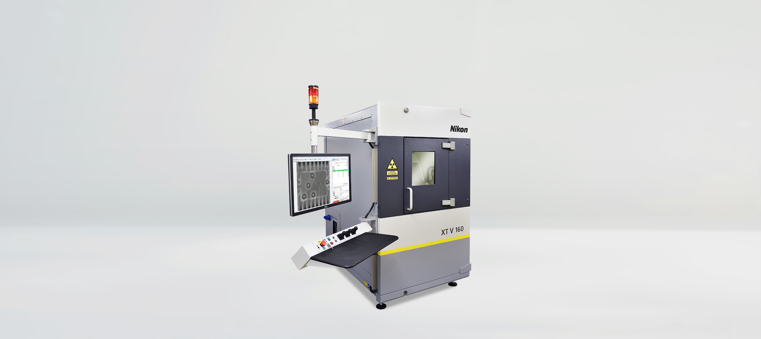
The X-ray CT scan of the entire specimen and the ROI of the skull were acquired an X-ray power of 16 and 7 Watts and a voxel resolution of 17 and 4 µm, respectively. Both scans were acquired using a Nikon XT H 225, which houses a Nikon 225 kV microfocus X-ray source fitted with a tungsten reflection target (transmission and rotating targets also available), coupled with a Varex 4343-CT flat panel detector. For these scans the detector acquired 4476 and 4424 projections at an exposure of 250 and 2829 ms, for the entire specimen and ROI skull scans, respectively.
X-ray CT data was reconstructed using a modified filtered back projection algorithm in Nikon in-house reconstruction software CT Pro 3D and rendered using the isorender and Wall Thickness Analysis module in Volume Graphics Studio Max 3.4.